All published articles of this journal are available on ScienceDirect.

Role of Ferroptosis in Diabetic Foot Ulcers: Pathophysiological Mechanisms, Biomarkers, and Therapeutic Potencies
 iD
Ali Golchin3 , 4 , *
iD
Authors Info & Affiliations
Abstract
Diabetic foot ulcer (DFU) is one of the major complications of diabetes mellitus and often results in persistent non-healing lesions, infections, and amputations. Recently, an iron-dependent form of programmed cell death, ferroptosis, has been shown to have an essential role in the poor regeneration of DFUs. This mini-review aims to study the role of ferroptosis in the pathophysiology of diabetic wounds, clinical biomarkers, and potential treatments by targeting ferroptotic pathways. Hence, PubMed-indexed studies on ferroptosis in relation to diabetic wound healing, including in vitro, animal, and clinical studies, were systematically reviewed. The research was evaluated based on molecular mechanisms, biomarkers, and preclinical and clinical evidence. Ferroptosis inhibits wound healing by promoting lipid peroxidation, oxidative stress, and cell death in fibroblasts, keratinocytes, and endothelial cells. Wound severity and healing are associated with tissue and circulation biomarkers such as Glutathione Peroxidase 4 (GPX4), Solute Carrier Family 7 Member 11 (SLC7A11), Acyl-CoA Synthetase Long-Chain Family Member 4 (ACSL4), Malondialdehyde (MDA), and 4-Hydroxynonenal (4-HNE). Several treatments, including small-molecule ferroptosis inhibitors, iron chelators, and extracellular vesicles from mesenchymal stem cells, have demonstrated therapeutic benefits in preclinical studies (in vitro and animal models) and early-phase clinical assessments, although no randomized controlled trials are currently available. Inhibition of ferroptosis is a promising and rational therapeutic approach to improve DFU healing. It is essential to normalize these biomarkers, optimize therapeutic regimens, and perform extensive, robust clinical trials to translate the preclinical findings for chronic diabetic wounds into effective patient treatments.
1. INTRODUCTION
Diabetic foot ulcers (DFUs) are a major and serious complication of diabetes mellitus, resulting in significant morbidity, excessive healthcare costs, and an increased risk of lower-limb amputation. Diabetic wound healing is compromised due to a complex interaction of factors including peripheral neuropathy, vascular insufficiency, chronic inflammation, oxidative stress, and dysfunction of the cellular machinery [1]. The molecular pathways underlying poor healing of DFUs are unclear, whereas the common process of wound healing is well documented [2]. Ferroptosis, a regulated, iron-dependent form of cell death, has recently emerged as a potential mediator of tissue injury in diabetes. This process is associated with increased lipid peroxidation and the accumulation of Reactive Oxygen Species (ROS), which cause cellular membrane damage and cytotoxicity [3]. Ferroptosis is induced by the metabolic disorder of GPX4 (SLC7A11 and disordered iron balance, unlike apoptosis or necrosis. Long-term hyperglycemia, mitochondrial dysfunction, and an increased labile iron pool create a microenvironment conducive to ferroptotic injury in diabetic tissues [4, 5]. In vitro and animal studies have demonstrated that ferroptosis impairs the functionality of fibroblasts, keratinocytes, and endothelial cells, which are essential to tissue regeneration, angiogenesis, and extracellular matrix (ECM), among other processes [6]. Human observational studies have shown that elevated levels of ferroptosis-related biomarkers are associated with poor wound healing in DFU patients [7]. These data elucidate the mechanistic link between oxidative stress, ferroptotic cell death, and chronic non-healing ulcers [4, 7]. While the field is under active research, it is still in an early stage. Much of the research to date has been done in vitro and in animal models. However, translation to human DFU pathophysiology is yet to be established. In addition, ferroptosis inhibitors, including ferrostatin-1 (Fer-1), liproxstatin-1 (Lip-1), and deferoxamine (DFO), have demonstrated promise in animal models, but their clinical efficacy and safety remain to be determined, as no human trials have been completed [8].
This review aims to gather relevant research on ferroptosis related to DFUs to provide important insights and effective strategies to improve patient outcomes in DFU treatment. The basic molecular mechanisms, the experimental and clinical research, and the available treatments are discussed. Identifying existing knowledge gaps can help guide future research and improve the management of diabetic wounds in the clinic.
Ferroptosis in diabetic wound healing has been studied in numerous review articles. But most of them mainly focused on either the molecular mechanisms or the therapeutic possibilities individually. This review provides a systematic and integrated investigation of ferroptosis in DFUs, including its molecular regulatory networks, ferroptosis-associated biomarkers, in vitro and animal experimental findings, new clinical insights, and current treatments. Moreover, the analysis thoroughly considers the limitations of existing evidence, discusses the difficulties in translating preclinical findings into clinical practice, and identifies potential future research directions, such as multi-omics approaches, nanotechnology-based therapeutics, and precision medicine. The current study brings these disciplines together into a coherent framework and provides a more clinically relevant perspective for researchers and clinicians involved in the treatment of DFUs.
1.1. Literature Search Strategy
This narrative review was conducted by performing an extensive literature search across the PubMed/MEDLINE, Scopus, Web of Science, and Google Scholar databases. Publications up to February 2026 related to the topic were collected by combining several of the following terms: “ferroptosis”, “DFUs”, “diabetic wound healing”, “iron metabolism”, “lipid peroxidation”, “oxidative stress”, “GPX4”, “SLC7A11”, “Nrf2”, “ACSL4”, “ferritinophagy”, and “ROS”. The search methodology was enhanced through the use of Boolean operators (AND/OR). Further research was identified by manual screening of relevant articles in reference lists. We included research articles in English, experimental investigations, observational human studies, clinical trials, and high-quality review articles. Publications lacking sufficient scientific evidence, duplicate articles, conference abstracts without full text, and non-English literature were excluded. Selected studies were critically reviewed for their relevance to molecular mechanisms of ferroptosis, biomarkers, preclinical data, clinical observations, and treatment options in DFUs.
1.2. Pathological Mechanism of Ferroptosis in DFUs
Persistent hyperglycaemia, inadequate perfusion, chronic inflammation, and oxidative stress create a milieu for the development of DFUs. These conditions collectively predispose reparative cells to ferroptosis and result in delayed wound healing [2, 7, 9]. Therefore, the importance of ferroptosis in DFUs has to be assessed by understanding the exact pathological mechanisms of ferroptosis.
1.3. Cellular Mechanisms of Ferroptosis
Ferroptosis is a form of programmed cell death that is iron-dependent, characterized by the accumulation of lipid peroxides and ROS, resulting in membrane disintegration and cell death in diabetic wounds [2, 9]. Ferroptosis is closely related to the disruption of key antioxidant pathways, particularly GPX4 and the cystine/glutamate antiporter SLC7A11, which are different from apoptosis or classic necrosis, resulting in impaired glutathione (GSH) production, reduced detoxification of lipid peroxides, and increased susceptibility of diabetic ulcer tissue to oxidative damage [2, 5, 9].
Iron-dependent processes, especially the Fenton reaction, produce highly reactive hydroxyl radicals when ferrous iron (Fe2+) reacts with hydrogen peroxide, resulting in increased intracellular oxidative stress in diabetic ulcers and constituting a major source of ROS and free radicals in ferroptosis [2, 10]. These radicals attack polyunsaturated fatty acids in membrane phospholipids, resulting in lipid peroxidation and compromised membrane integrity, which in turn impedes the efficient healing of diabetic ulcers by favoring regulated cellular death pathways such as ferroptosis [2, 9, 10]. The chronic inflammatory milieu of DFUs sustains ROS production and lipid peroxidation because of disrupted iron homeostasis, characterized by elevated labile iron, modified ferritin storage and imbalanced iron absorption and excretion [2, 10].
The process of lipid peroxidation during ferroptosis is modulated by the availability of ROS and the enzymes involved in membrane phospholipid modification that control the levels of polyunsaturated fatty acids (PUFAs), such as ACSL4 and lysophosphatidylcholine acyltransferase 3 (LPCAT3) [2, 9]. These enzymes promote the incorporation of PUFA-containing phospholipids into cell membranes, which are particularly vulnerable substrates for oxidation. Reduced GPX4 activity leads to both enzymatic and non-enzymatic peroxidation of PUFA-phospholipids, producing lipid hydroperoxides and cytotoxic aldehydes that impair membrane integrity and function [2, 9]. In the setting of DFUs, oxidative stress due to hyperglycaemia, accumulation of advanced glycation end products, and persistent low-grade inflammation downregulate the expression of SLC7A11 and GPX4, thereby impairing the antioxidant defence and leading to susceptibility of keratinocytes, fibroblasts, and endothelial cells in the wound bed to ferroptosis [2, 5, 9].
Besides these basic metabolic disturbances, there is a significant interaction among ferroptosis, the SIRT1/Nrf2/p62 signaling pathway, and autophagy, which seems particularly relevant in DFUs [10]. Activation of SIRT1 promotes deacetylation and nuclear translocation of Nrf2, which induces transcription of antioxidant and cytoprotective genes, including those involved in GSH production and iron sequestration, resulting in decreased lipid peroxidation and ferroptotic damage in the wound microenvironment [5, 10]. At the same time, selective autophagy mediated by p62 regulates the degradation of Keap1 and sustains the activation of Nrf2, while the autophagy-mediated degradation of ferritin and damaged mitochondria dynamically modulates the labile iron pool and mitochondrial ROS generation, linking autophagic flux to the susceptibility of ferroptosis in diabetic foot tissue [2, 10]. In animal models of DFUs, pharmacological or genetic activation of the SIRT1/Nrf2/p62 pathway has been shown to rescue oxidative stress, restore autophagy, inhibit excessive ferroptosis, and subsequently enhance granulation tissue formation, angiogenesis, and re-epithelialization, suggesting this autophagy–ferroptosis crosstalk as a feasible therapeutic target to improve diabetic wound healing [2, 5, 9, 10]. The main mechanisms of ferroptosis are shown in Fig. (1), including iron metabolism, lipid peroxidation, and blockade of the antioxidant system.
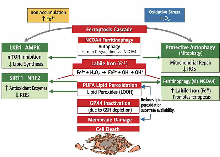
Schematic overview of the main mechanisms of Ferroptosis; Schematic representation of the key molecular events involved in ferroptosis initiation and progression. Iron accumulation increases the intracellular ferrous iron (Fe2+) pool, while oxidative stress, including hydrogen peroxide (H2O2) generation, promotes iron-dependent lipid peroxidation. Excess labile iron participates in Fenton chemistry, converting hydrogen peroxide into reactive hydroxyl radicals (•OH), which enhance lipid oxidation and cellular damage.
Abbreviations: AMPK: adenosine monophosphate-activated protein kinase, GPX4: glutathione peroxidase 4, GSH: glutathione, LKB1: liver kinase B1, LOOH: lipid hydroperoxides, mTOR: mechanistic target of rapamycin, NCOA4: nuclear receptor coactivator 4, NRF2: nuclear factor erythroid 2–related factor 2, PUFA: polyunsaturated fatty acid, ROS: reactive oxygen species, SIRT1: silent information regulator 1.
1.4. Ferroptosis-related Biomarkers in DFUs
The biomarkers linked to ferroptosis could provide essential data on the activation of ferroptosis mechanisms and may mirror critical pathological events, such as iron dysregulation, lipid peroxidation, and antioxidant defense dysfunction. Table 1 summarizes the ferroptosis-related biomarkers in DFU. Ferroptosis biomarkers are mainly identified by changes in critical regulatory pathways such as the GSH/GPX4 axis, SLC7A11-mediated cystine transport, iron metabolism, and lipid peroxidation mechanisms [11, 12].
| Biomarker | Biological Role | Evidence | Clinical Significance | Therapeutic Potential | Refs. |
|---|---|---|---|---|---|
| GPX4 | Prevents lipid peroxidation | Cell + Animal +Limited Human | Potential diagnostic/Prognostic marker | GPX4 activation may reduce ferroptosis | [5, 11] |
| SLC7A11 | Cystine uptake and GSH synthesis | Cell + Animal | Reflects antioxidant defense | Target to restore redox balance | [5, 11] |
| ACSL4 | PUFA metabolism; promotes ferroptosis | Cell + Animal | Associated with ferroptosis susceptibility | ACSL4 inhibition may protect tissues | [11, 13] |
| Nrf2 | Master regulator of antioxidant genes | Cell + Animal + Limited Human | Indicator of oxidative stress response | Activation may suppress ferroptosis | [5] |
| Ferritin | Iron storage | Human + Experimental | Reflects iron overload | Iron chelation strategies | [5, 11] |
| Transferrin Receptor (TFR1) | Cellular iron uptake | Experimental | Indicator of iron metabolism | Potential therapeutic target | [5, 11] |
| MDA | Lipid peroxidation product | Human + Experimental | Marker of oxidative damage | Useful for disease monitoring | [11, 13] |
| 4-HNE | Lipid peroxidation aldehyde | Experimental | Oxidative stress biomarker | Research biomarker | [11] |
Several molecules associated with ferroptosis, such as GPX4, SLC7A11, ACSL4, MDA, 4-HNE, and proteins related to iron metabolism, have been proposed as potential markers of ferroptotic activity based on in vitro, animal, and limited human observational studies [12, 13]. Reduced GPX4 and SLC7A11 expression points to impaired antioxidant defense, while increased ACSL4 levels and build-up of lipid peroxidation products such as MDA and 4-HNE suggest heightened ferroptotic signalling [12, 13].
In diabetic wounds, dysregulation of ferroptosis-related markers may impair cell function and delay tissue regeneration. Recent in vitro and human tissue studies suggest that diabetic wound tissues and keratinocytes exhibit features of ferroptosis, such as elevated ACSL4, elevated MDA and 4-HNE levels, and altered regulation of pathways related to ferroptosis, suggesting that these molecules could serve as potential markers for ferroptosis activity in diabetic wound healing [13]. However, further studies are needed to validate their clinical utility as diagnostic or prognostic biomarkers in DFUs.
1.5. Ferroptosis Inducers in Diabetic Wounds
Ferroptosis inducers are agents or conditions that induce ferroptosis. Indeed, they are the main drivers of the pathophysiological process of ferroptosis in DFUs. There are several diabetes-related changes that favor the occurrence of ferroptosis in DFUs.
1.5.1. Long-term hyperglycemia
The persistent high glucose level promotes ROS generation, oxidative stress, and reduces the threshold of the white line for ferroptosis [2, 4].
1.6. Chronic Inflammation and Poor Perfusion
Infiltrating inflammatory cells produce ROS and consume nutrients, which pose additional barriers to reparative cells [5, 14].
Regulatory RNAs and gene networks have been attributed to the development of non-healing diabetic wounds, including CircRNAs, miRNAs, and ferroptosis-related mechanisms [15, 16].
Fibroblast, keratinocyte, and endothelial cell ferroptosis strongly hinders tissue regeneration, particularly in diabetic injuries. A principal driver of ferroptosis is the accumulation of iron, which is often observed in diabetes. Excess free iron increases lipid peroxidation via the Fenton reaction, resulting in cell death and dysfunction of reparative cells [2, 6].
- Fibroblasts: Ferroptotic fibroblasts showed reduced synthesis of type I collagen and ECM, which hampered the formation of granulation tissue [6, 17].
- Endothelial cells: Ferroptosis inhibits angiogenesis and vascular development, which limits the supply of oxygen and nutrients to the wound site [6].
- Keratinocytes: Decreased keratinocyte proliferation and viability impair epithelialization and wound healing [2, 6].
- In vitro studies have shown that GPX4 and SLC7A11 are expressed at lower levels, while ACSL4 is expressed at higher levels under high glucose conditions, revealing the onset of ferroptosis [2, 6]. Importantly, in vitro experiments have demonstrated that ferroptosis inhibition by Fer-1 or DFO rescues cell viability, motility, and angiogenic activity [6, 17]. These findings highlight iron-mediated ferroptosis as a major contributor to impaired granulation tissue, reduced angiogenesis, and delayed wound healing in DFUs, and provide potential therapeutic approaches for promoting wound healing [2, 6].
1.7. Clinical and Translational Evidence
The importance of ferroptosis in the pathophysiology of DFUs has been demonstrated increasingly through bioinformatic, experimental, translational, and clinical studies (Table 2) [10, 14]. Bioinformatic analyses of human tissue datasets from DFU patients have demonstrated alterations in multiple ferroptosis-related genes (e.g decreased GPX4 and increased ACSL4) and changes in regulatory networks that are implicated in the defective wound healing process [10]. Consistent with these observations, studies on senescent fibroblasts have identified impaired NCOA4-dependent ferritinophagy leading to dysfunctional ferroptosis and retarded wound healing [14].
| Study Type | Model / Samples | Main Findings | Ferroptosis Markers / Pathways | Relevance to DFU Healing | Refs. |
|---|---|---|---|---|---|
| Bioinformatics + Clinical | DFU patient tissue datasets | Identified key ferroptosis-related genes and regulatory networks involved in DFU pathology | GPX4 ↓, ACSL4 ↑, other gene signatures | Supports ferroptosis involvement in DFU impairment | [10] |
| Cellular Experimental Study | Senescent fibroblasts | Detected defective NCOA4-dependent ferritinophagy and ferroptosis in aged fibroblasts | NCOA4 pathway dysregulation | Defective ferroptosis contributes to delayed diabetic wound healing | [14] |
| Network Pharmacology + In vitro + In vivo | DFU wound models + computational | Dracorhodin improves DFU healing through activation of the Nrf2 axis and ferroptosis suppression | Nrf2 activation → decreased lipid peroxidation | Reduced ferroptosis accelerates DFU wound closure | [16] |
| In vitro + In vivo Experimental | DFU animal model + cell culture | Metrnl mitigates ferroptosis by preventing mitochondrial damage via LKB1/AMPK | LKB1/AMPK pathway; reduced mitochondrial ROS | Inhibition of ferroptosis improves DFU healing outcome | [18] |
| Cell therapy + in vivo | DFU wound models | Transplanted FGF21-ADSCs reduced oxidative stress and ferroptosis in wound tissue, enhanced angiogenesis, and accelerated wound closure in diabetic models. | SIRT1 → NRF2 → ↑ GPX4; decreased lipid peroxidation & inflammatory signaling | suggests cell-based therapy as a viable approach to reduce ferroptosis and promote wound repair in diabetes. | [19] |
| In vitro (HUVEC under high glucose) + in vivo | DFU wound models | Resveratrol treatment increased cell survival, reduced ROS and lipid peroxidation, and enhanced angiogenesis and wound healing in diabetic models. | ↑ GPX4, ↑ SLC7A11; ↓ ROS, ↓ lipid peroxidation — via PI3K-AKT-Nrf2 pathway | Indicates that natural antioxidant compounds can mitigate ferroptosis and improve healing — potential low-cost adjunct therapy. | [20] |
| In vitro (endothelial cells under high glucose) + in vivo | DFU wound model | Artemisinin activated Nrf2/GPX4, decreased intracellular iron and lipid ROS, improved endothelial function and wound healing outcomes. | ↑ Nrf2 → ↑ GPX4; ↓ iron overload, ↓ lipid peroxidation | Adds evidence for small-molecule interventions targeting ferroptosis as a therapeutic strategy in DFU. | [21] |
| Clinical observational | DFU patients | Higher serum NRF2 levels correlated with better wound healing outcomes and lower inflammatory markers. | NRF2 (serum) as a predictive biomarker | Provides clinical evidence for NRF2’s prognostic value — potential for using ferroptosis-related biomarkers in DFU management. | [22] |
Furthermore, in vitro and animal studies also support the clinical significance of targeting ferroptosis in DFUs. Compounds such as dracorhodin have been shown to promote wound healing in diabetic models through activation of the Nrf2 pathway and inhibition of lipid peroxidation [16]. Protein Meteorin-like (Metrnl) also alleviates ferroptosis-associated injury by maintaining mitochondrial integrity and stimulating the LKB1/AMPK signaling pathway [18]. Cellular therapy holds promise; FGF21-modified adipose-derived stem cells (ADSCs) activate the SIRT1–NRF2–GPX4 pathway to decrease oxidative stress, inflammation, and ferroptotic cell death in the wound environment, promoting angiogenesis and wound repair [19].
Natural substances also provide evidence further supporting the translational viability of therapies that modulate ferroptosis. Resveratrol and artemisitene were shown to promote wound healing in DFU models by reducing intracellular iron accumulation, lipid ROS, and oxidative damage through Nrf2 activation and GPX4 upregulation [20, 21]. These results demonstrate the possibility of using inexpensive additional treatments with antioxidants. Importantly, human observational data support the significance of ferroptosis-related pathways in DFU patients. Increased serum NRF2 levels in DFU patients are associated with better prognosis, less inflammation, and better healing outcomes, suggesting a potential role of NRF2 as a biomarker for DFU prognosis [22]. Altogether, the data indicate that ferroptosis is a promising target for the development of advanced, mechanism-directed therapies and a contributor to the pathophysiology of DFU.
1.8. Experimental and Animal Models on Ferroptosis in DFUs
Several preclinical studies (in vitro and animal models) have been conducted to investigate the role of ferroptosis in diabetic wound healing (Table 3) [3, 5]. These studies provide additional mechanistic insight and suggest possible therapeutic targets (Fig. 2) [3, 5]. Several cell culture studies have also mimicked the diabetic wound microenvironment by exposing fibroblasts, endothelial cells, and keratinocytes to high glucose levels, all evidencing ferroptosis-like events [3]. For instance, fibroblasts and endothelial cells augmented with glucose exhibit downregulated GPX4 and SLC7A11, elevated lipid peroxidation byproducts (MDA, 4HNE), upregulated ACSL4, and reduced survival and migratory function; the ferroptosis inhibitor Fer-1 partially counteracts these effects and recovers partial functionality [3, 5]. Preliminary studies demonstrate that NETosis (Neutrophil Extracellular Trap formation is a process by which neutrophils release DNA and antimicrobial proteins to trap pathogens) in diabetic-wound endothelial cells initiates ferroptosis through inhibition of the PI3K/AKT pathway, resulting in a reduction of angiogenic potential [23]. The in vitro data support a potential link between increased glucose stress, induction of ferroptosis, and defective cellular function in wound repair [3].
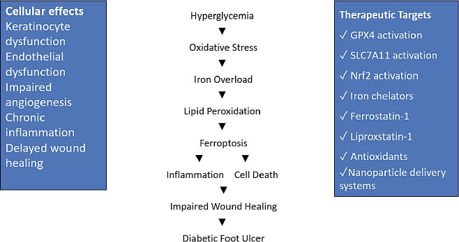
Overview of ferroptosis-mediated molecular mechanisms, therapeutic targets, and potential interventions in DFUs. Schematic illustration depicting the pathological cascade linking hyperglycemia-induced metabolic disturbances to ferroptosis-mediated impairment of diabetic wound healing;
Abbreviations: DFU: diabetic foot ulcer, GPX4: glutathione peroxidase 4, SLC7A11: solute carrier family 7 member 11, Nrf2: nuclear factor erythroid 2–related factor 2.
| Model Type | Experimental Details | Ferroptosis-Related Findings | Outcome on Wound Healing / Cell Function | Refs. |
|---|---|---|---|---|
| In vitro (fibroblasts & endothelial cells under high glucose) | PRP treatment of HSF & EA.HY926 cells cultured in a high-glucose environment | ↓ ACSL4; ↑ GPX4 & SLC7A11; ↓ ROS & MDA | Improved cell proliferation & migration; reduced inflammation; restored redox balance | [9] |
| In vivo (STZ-induced diabetic rats) | PRP injected into diabetic skin wounds | ↓ oxidative stress markers; ↑ antioxidant activity | Accelerated wound closure; reduced inflammatory markers | [9] |
| In vitro (fibroblasts, endothelial cells, keratinocytes under high glucose) | High-glucose exposure modeling diabetic microenvironment | ↓ GPX4, ↓ SLC7A11; ↑ lipid peroxidation (MDA, 4-HNE); ↑ ACSL4; reduced survival & migration | Fer-1 partially reverses ferroptosis markers and restores cell function | [3] |
| In vitro (endothelial cells) | NETosis-induced ferroptosis under diabetic conditions | NETosis inhibits PI3K/AKT → triggers ferroptosis | Reduced angiogenic potential; EVs from MSCs reduce ferroptosis and improve function | [23] |
| In vivo (STZ-diabetic rats) | Topical Fer-1 | ↓ inflammatory & oxidative markers | Accelerated wound healing; ↑ CD31/VEGF | [5] |
| In vivo (mouse model of diabetic foot ulcer) | Overexpression of Metrnl | Activates LKB1/AMPK; suppresses mitochondrial injury and ferroptosis | Promoted wound healing | [18] |
| In vivo (diabetic mice) | Modulation of ferritinophagy via NCOA4 expression | SICLE resistant to ferritinophagy; NCOA4 ↑ ferroptotic sensitivity | Improved wound healing with NCOA4 overexpression | [14] |
Topical application of ferroptosis modulators in diabetic murine models led to improved wound healing [5, 9]. For example, topical administration of Fer-1 in streptozotocin-induced diabetic rats resulted in improved wound healing, increased CD31/VEGF levels, and decreased expression of inflammation and oxidative markers in comparison to untreated controls [5]. Increased expression of the secreted Metrnl activated the LKB1/AMPK pathway to attenuate mitochondrial injury and ferroptosis and promote wound healing in a murine model of DFUs [18].
Aging fibroblasts were resistant to ferritinophagy, while overexpression of nuclear receptor co-activator 4 (NCOA4) enhanced ferroptosis susceptibility and promoted wound healing in diabetic mice [14]. These animal model findings suggest that modulation of ferroptosis could be a therapeutic approach to enhance impaired wound healing in diabetes [3, 5].
1.9. Translational Insights from Pre-clinical Studies to Research Gaps
In animal experiments, prolonged healing has been associated with elevated levels of ferroptosis markers. Common features to be observed in diabetic wound tissue with diminished antioxidant defense are iron accumulation, increased ROS, and lipid peroxidation [7]. For instance, both intracellular free iron and 4-HNE were shown to be significant predictors of reduced rates of wound healing in rodent DFU models [7, 8]. Some regenerative therapies have been reported to influence endothelial cell ferroptosis. For example, in diabetic mouse models, mesenchymal stem cell-derived extracellular vesicles (MSC-EVs) can inhibit endothelial cell ferroptosis, promote angiogenesis, and accelerate wound healing [24]. Altogether, these findings indicate that the use of therapeutic strategies aimed at inhibiting ferroptosis may be applicable with regard to diabetic wound healing.
Although pre-clinical results look promising, a number of questions remain unanswered. The long-term safety and efficacy of ferroptosis inhibitors in diabetic wound models remain unclear. The majority of in vivo investigations are short-acting, and rodent models are mainly being used; large-animal or humanized models still do not exist. Moreover, there is scarce biomarker validation for ferroptosis in human DFUs. The crosstalk between ferroptosis and other death subtypes (e.g., pyroptosis, autophagy) in DFU needs to be further explored.
1.10. Clinical Indication and Biomarkers of Ferroptosis in the DFUs
As mentioned above, DFU is an important clinical problem because persistent failure of the wound to heal is associated with increasing risk of infection, amputation, and morbidity. Although animal studies have shown an implication of ferroptosis in retarded wound healing, human evidence is now available to confirm it as a clinically relevant phenomenon. Human tissue biopsy studies from DFUs have demonstrated upregulation of ferroptosis markers, including reduced GPX4, downregulation of SLC7A11, and upregulation of ACSL4, which contribute to impaired regeneration and oxidative stress in wound beds [2, 9, 10].
In human DFU tissue samples, ferroptosis-related lipid peroxidation products such as MDA and 4-HNE are increased compared with non-diabetic control skin, indicating an excessive iron-catalyzed ROS accumulation [2, 5]. Most notably, their abundance has been inversely related to wound closure rates and positively related to ulcer severity and risk of infection, thereby suggesting a potential role in prognostication [10, 18]. Circulating biomarkers of ferroptosis have also been investigated in addition to tissue-specific markers. In human observational studies, serum ferritin and transferrin saturation, and LPI are higher in patients with chronic DFUs, whereas systemic antioxidants such as GSH and GPX4 activity are decreased [4, 16]. These results indicate that local tissue and systemic ferroptotic events may play a role in the outcomes of wounds as well as serve as a non-invasive window for clinical monitoring. Accordingly, recent transcriptomic and proteomic studies conducted with human DFU tissues have contributed to further defining the molecular mechanisms of ferroptosis in diabetic wounds (Fig. 2). Disruptions in Nrf2, p53, and NF-κB signalling have been shown to contribute to the lipid peroxidation and ferroptotic cell death that is observed, together with increased levels of iron import proteins (e.g., transferrin receptor 1), which results in further accumulation of intracellular iron [8, 15, 18].
Furthermore, integrative analysis of these data also identifies potential therapeutic targets or drugs, such as GPX4 activators and iron chelators, for ameliorating cell and tissue damage by ferroptosis in DFU patients.
Thus, upon analysis of clinical outcomes, correlative studies further demonstrate the association between ferroptosis markers and prognosis. Patients with high levels of MDA, 4-HNE, and ACSL4 in the tissue or serum often have delayed healing, secondary infection occurrence, and surgical requirement [14, 23]. Conversely, Treatments that replenish antioxidant potential or inhibit lipid peroxidation, like the use of Fer-1 analogs topically or stem cell-derived exosomal-based therapies, have been reported with better healing rates in small-scale clinical studies, although these remain preliminary and require confirmation in larger trials [4, 9, 24]. However, there are still great challenges in translating ferroptosis from preclinical to clinical applications.
Limitations include small patient cohorts, heterogeneity in biomarker assays, and the absence of standardized thresholds for tissue ferroptosis activity in DFU. In addition, there is a lack of comprehensive understanding of the crosstalk between ferroptosis and other forms of regulated cell death, including apoptosis, pyroptosis, and necroptosis, in chronic diabetic wounds [7]. It is important to address these knowledge gaps to develop targeted and safe interventions for human populations.
Collectively, the clinical evidence summarized above suggests that ferroptosis plays an important role in the evolution of DFU and treatment resistance. Tissue-specific and systemic biomarkers are associated with wound severity and healing and may provide opportunities for patient stratification and prognostication. Integrating molecular, histological, and systemic data related to ferroptosis represents a promising approach for identifying novel therapeutic strategies targeting the ferroptotic cascade. However, efforts should focus on overcoming current limitations and translating these findings into clinical practice.
1.11. Therapeutic Methods for Targeting Ferroptosis in DFUs
Each therapeutic intervention acts through a variety of molecular pathways. Either artificially activating GPX4 or increasing the levels of GSH directly inhibits lipid peroxidation, or functionally upregulating Nrf2 signaling that enhances antioxidant enzyme transcription to decrease susceptibility to ferroptosis [24]. Inhibition of ACSL4, the key enzyme that incorporates polyunsaturated fatty acids into phospholipids, reduces lipid peroxidation and membrane susceptibility [24]. Furthermore, modulation of transferrin receptor 1 and ferritin expression to control iron homeostasis regulation is involved in the wound healing process, suggesting that multi-target interventions would also be required to successfully alleviate ferroptotic cell death in diabetic wounds [24].
However, inhibition of ferroptosis is a promising strategy for accelerating wound healing in DFUs by connecting basic molecular studies to clinical therapies [16, 24]. The overall burden of ferroptotic cell death in fibroblasts, keratinocytes, and endothelial cells leads to impaired angiogenesis, ECM production, and delayed re-epithelialization; consequently, it is a logical target for both pharmacological and cell-based treatments [24].
The majority of therapeutic studies have been performed in vitro or in animal models, showing that ferroptosis inhibition is a reliable and beneficial strategy to improve wound healing [16, 18, 25]. For example, diabetic animal models receiving Fer-1 or Exo-MSC (Exosomes from Mesenchymal Stem Cells) show faster wound closure rates, increased angiogenesis and decreased oxidative stress markers [25]. Preliminary clinical trials have started to assess the safety and efficacy of antioxidant therapy, as well as iron chelation in patients with chronic DFUs; however, targeted ferroptosis induction or rescue has been limited in humans, and no randomized controlled trials have been completed [24]. Altogether, these findings indicate that regulation of ferroptosis could be a new adjunctive approach for DFU treatment [16, 18, 25]. Table 4 summarizes therapeutic strategies targeting ferroptosis in DFU.
| Therapeutic Method | Molecular Target / Pathway | Mechanism of Action | Observed Effects / Outcome | Model / Context | Ref. |
|---|---|---|---|---|---|
| GPX4 activation / GSH increase | GPX4 | Inhibits lipid peroxidation, protects cells from ferroptosis | Reduced cell death, enhanced wound healing | In vitro / DFU model | [16] |
| Nrf2 signaling upregulation | Nrf2 | Enhances antioxidant enzyme transcription, reduces susceptibility to ferroptosis | Decreased oxidative stress, improved fibroblast and keratinocyte survival | In vitro / DFU model | [16] |
| ACSL4 inhibition | ACSL4 enzyme | Direct inhibition of ACSL4 → reduced incorporation of ECMs into phospholipids → decreased lipid peroxidation and membrane susceptibility to ferroptosis | Inhibition of ferroptotic cell death, reduced lipid peroxidation | In vitro / in vivo general ferroptosis models (not DFU-specific) | [24] |
| Iron homeostasis modulation | Transferrin receptor 1, Ferritin | Regulates iron levels → reduces ferroptosis susceptibility | Supports wound healing, reduces ferroptosis | Senescent fibroblasts / DFU model | [26] |
| Pharmacological ferroptosis inhibitors (Fer-1, Exo-MSC) | Multiple pathways | Direct ferroptosis inhibition | Faster wound closure, increased angiogenesis, decreased oxidative stress | Animal models / exosomal treatment | [25] |
| Antioxidant therapy / Iron chelation | ROS, iron overload | Reduce oxidative stress and iron-induced damage | Safety and efficacy being assessed in humans | Preliminary clinical trials / DFU patients | [18] |
1.12. Pharmacological Inhibitors of Ferroptosis
A number of small-molecule inhibitors have been studied in diabetic wound models.
In vitro studies have demonstrated that Fer-1 and Lip-1 are effective in inhibiting lipid peroxidation, reducing ROS accumulation, rescuing cell viability, and enhancing fibroblast migration. In streptozotocin-induced diabetic mouse models, topical Fer-1 promotes wound healing by accelerating the process of wound closure, increasing Vascular Endothelial Growth Factor (VEGF) expression, and reducing levels of inflammatory cytokines in the wound beds [2, 4, 24].
Iron chelation with DFO attenuates the accumulation of LMF (Low Molecular Fraction, a small molecular-weight component associated with lipid peroxidation, which accumulates in chronic wounds) and, in turn, reduces ROS-mediated lipid peroxidation and enhances granulation tissue formation [23]. These findings offer strong evidence for the effectiveness of ferroptosis modulators in preclinical DFU models.
1.13. Cell-Based and Regenerative Therapies
Exosomes derived from MSCs and other cell-based therapies have been reported to play roles in inhibiting ferroptosis, as well as in promoting tissue repair [27, 28]. MSC EVs transmit microRNAs (such as miR-548ai, miR-660, miR-1275, miR-23a-3p) and proteins to promote GPX4 expression, activate the Nrf2 antioxidant pathway, and alleviate intracellular iron-mediated oxidative stress in endothelial cells and fibroblasts [26, 29, 30]. In diabetic mouse models, MSC-derived exosomes promoted wound healing by decreasing wound closure time, enhancing collagen formation, and vascular regeneration [8]. These results illustrate the translational potential of cell-based therapies in regulating ferroptosis for the treatment of DFUs. However, regenerative medicine offers potential strategies for enhancing chronic wound healing by the utilization of therapeutic medicines and adjuvants, especially those derived from cells and tissues [31].
1.14. Translational Challenges and Future Directions
Despite substantial progress in our understanding of the role of ferroptosis in DFUs, several major obstacles remain to its clinical application. Most of the available evidence is based on in vitro and animal studies, and few large-scale clinical trials have been completed to date. The complex pathophysiology of human DFUs, including persistent inflammation, peripheral nerve damage, inadequate blood flow, infection, and metabolic abnormalities, cannot be adequately replicated in experimental models. In addition, many ferroptosis-related biomarkers such as GPX4, ACSL4, SLC7A11, and lipid peroxidation products have shown promising experimental results but lack standardized measurement methods, validated diagnostic criteria, and prospective clinical validation in human cohorts. Thus, their diagnostic and prognostic value in routine clinical practice is unclear.
Therapeutic modulation of ferroptosis has many challenges. As ferroptosis is involved in normal physiological functions, its indiscriminate inhibition may cause unexpected biological consequences. In addition, problems of drug delivery, tissue selectivity, long-term safety, and optimal timing of treatment remain unresolved. Therefore, large multicenter clinical trials are needed before ferroptosis-related biomarkers and treatment strategies can be incorporated into the routine care of diabetic foot ulcers.
Furthermore, the complicated and multifactorial pathophysiology of DFU, including impairment of angiogenesis, chronic inflammation, neuropathy, immune dysfunction, and oxidative stress, makes it difficult to determine how ferroptosis exactly fits within this intricate network. The involvement of oxidative stress and lipid peroxidation in the development of diabetic ulcers also contributes to the difficulty in identifying specific biomarkers and understanding the underlying mechanisms [2].
Another complication is the identification of sensitive and specific biomarkers. Most candidate markers associated with ferroptosis studied to date (e.g., lipid peroxidation products or iron-processing proteins) are not specific and may overlap with other oxidative stress or inflammatory pathways [2, 7, 10, 32]. The ideal dose, route of delivery, and nature of ferroptosis inhibition or cellular therapy remain unknown; these approaches are associated with concerns regarding off-target effects and potential disruption of essential iron and lipid metabolism homeostasis. Further studies are also needed to explore the relationship between ferroptosis and other regulated cell death (RCD) pathways, such as apoptosis, necroptosis, and pyroptosis. Evidence suggests that inhibition of one pathway may redirect cellular death toward alternative mechanisms, potentially affecting tissue injury outcomes [32].
Moreover, significant translational hurdles remain, including patient heterogeneity among individuals with type 1 and type 2 diabetes, differences in glycemic control, vascular limitations, comorbidities, and the lack of standardized large animal models. Ultimately, validation of the safety, efficacy, and clinical relevance of ferroptosis-targeted therapies will require well-designed translational studies and controlled clinical trials [13, 32, 33].
Future studies should focus on the development of novel therapeutic approaches, including specific ferroptosis inhibitors, iron chelators, and improved drug delivery systems such as hydrogels or nanoparticles suitable for chronic wound environments [34]. The studies described above raise the possibility that modulation of iron homeostasis and lipid peroxidation could promote wound healing and provide clinically significant benefits [33].
Personalized medicine may play an important role in this regard. Patient stratification and individualized therapeutic approaches could be achieved based on molecular profiling, transcriptomic signatures, and ferroptosis-related gene networks. For instance, key regulatory molecules such as ACSL4 and YTHDF2 (an m6A “reader” protein that recognizes methylated RNA and promotes mRNA decay, thereby regulating gene expression) have been implicated as potential molecular therapeutic targets in DFU [13].
Ultimately, further investigations should focus on translating mechanistic insights into clinically relevant diagnostic and therapeutic approaches. Multi-omics approaches, including genomics, transcriptomics, proteomics, metabolomics, and lipidomics, may provide a comprehensive understanding of the molecular networks involved in ferroptosis in DFUs. In addition, single-cell RNA sequencing may help identify cell-specific ferroptosis markers and clarify interactions among keratinocytes, fibroblasts, endothelial cells, macrophages, and immune cells during wound healing. Nanotechnology-based pharmaceutical delivery systems represent a promising strategy for delivering ferroptosis modulators to target sites while minimizing systemic side effects. Molecular biomarkers combined with clinical characteristics may enable personalized treatment strategies and improve patient stratification. Future multicenter clinical trials with rigorous design are required to validate ferroptosis-related biomarkers and evaluate the safety and efficacy of ferroptosis-targeted therapies in patients with DFU.
CRITICAL SUMMARY AND CONCLUSION
Ferroptosis has been recognized as an important mechanism that obstructs wound healing in DFUs due to altered iron metabolism, increased lipid peroxidation, oxidative stress, and inflammatory signaling. Accumulated in vitro and animal evidence suggests that targeting ferroptosis may be a promising therapeutic strategy. Several ferroptosis-related biomarkers have been identified in experimental and limited human observational studies, demonstrating diagnostic and prognostic potential. However, the current evidence is mostly preclinical, and significant hurdles need to be overcome before clinical routine application. Future studies should focus on extensive clinical validation, uniform evaluation of biomarkers, development of safe ferroptosis-targeted treatments, and integration of new technologies such as multi-omics and precision medicine. These initiatives may ultimately enable the translation of ferroptosis research into improved clinical outcomes in patients with diabetic foot ulcers.
AUTHORS’ CONTRIBUTIONS
The authors confirm their contributions to the paper as follows: A.G. and A. M.: Conceived the study, designed and supervised the review, and A. A., A.Z and F. F. conducted the study and wrote the manuscript. All the authors read and approved the final manuscript.
LIST OF ABBREVIATIONS
| DFU | = diabetic foot ulcer |
| GPX4 | = glutathione peroxidase 4 |
| SLC7A11 | = solute carrier family 7 member 11 |
| GSH | = glutathione |
| ACSL4 | = acyl-CoA synthetase long-chain family member 4 |
| MDA | = malondialdehyde |
| 4-HNE | = 4-hydroxynonenal |
| ROS | = reactive oxygen species |
| ECM | = extracellular matrix |
| DFO | = deferoxamine |
| Fer-1 | = ferrostatin-1 |
| Lip-1 | = liproxstatin-1 |
| NAC | = N-acetylcysteine |
| CoQ10 | = coenzyme Q10 |
| MSC | = mesenchymal stem cell |
| miRNA | = microRNA |
ACKNOWLEDGEMENTS
The authors acknowledge the utilization of AI-driven language help (QuillBot) peerless for grammatical improvements, stylistic enhancement, and designing figures.