All published articles of this journal are available on ScienceDirect.

Serum Endoglin as a Predictive Biomarker for Active Digital Ulcers in Systemic Sclerosis: A Case-Control Study
 iD
iD
Authors Info & Affiliations
Abstract
Introduction
This study aimed to evaluate soluble endoglin (sENG) in serum as a potential biomarker of active digital ulcers (DUs) in systemic sclerosis (SSc).
Methods
This single-center, cross-sectional case-control study included 60 SSc patients, comprising 30 with active DUs and 30 without active DUs, and 30 age- and sex-matched healthy controls. Serum sENG levels were measured using enzyme-linked immunosorbent assay.
Results
Median sENG levels were highest in SSc patients with active DUs, followed by those without active DUs and healthy controls: 0.54, 0.28, and 0.19 ng/mL, respectively. Patients with active DUs had significantly higher sENG levels than those without active DUs (p = 0.0007). Among SSc patients without active DUs, sENG levels did not differ significantly between those with previous but inactive DUs and those who had never had DUs (p = 0.344). In contrast, patients with active DUs had significantly higher sENG levels than both DU-naïve patients (p = 0.009) and patients with previous but inactive DUs (p < 0.05). ROC analysis showed that an sENG cut-off of 0.24 ng/mL differentiated SSc patients from healthy controls, with an AUC of 0.91, while a cut-off of 0.43 ng/mL discriminated SSc patients with active DUs from those without active DUs, with an AUC of 0.75. sENG levels were positively correlated with the number of active DUs (rho = 0.39, p = 0.002), but not with DUCAS-assessed DU severity (rho = -0.09, p = 0.65). Higher sENG levels were also associated with active/late nailfold capillaroscopy patterns and elevated systolic pulmonary arterial pressure.
Discussion
These findings suggest that serum sENG may reflect active DU-related microvascular injury rather than remote DU history alone. Its association with active ulcer burden, advanced nailfold capillaroscopy patterns, and elevated systolic pulmonary arterial pressure supports its potential role as an accessible adjunctive biomarker of active vascular involvement in SSc, although longitudinal validation is required.
Conclusion
Serum sENG may serve as a potential biomarker of active DU burden and ongoing microvascular injury in SSc.
1. INTRODUCTION
Systemic sclerosis (SSc) is a complex multisystem autoimmune connective tissue disease characterized by immune dysregulation, progressive fibrosis, and widespread microvascular injury [1]. Vascular involvement is considered an early and central event in SSc pathogenesis, often preceding overt skin or visceral fibrosis and manifesting as Raynaud’s phenomenon, nailfold capillaroscopy abnormalities, ischemic digital ulcers, pulmonary arterial hypertension, and, in severe cases, scleroderma renal crisis [2]. Endothelial activation, reduced nitric oxide bioavailability, excessive vasoconstrictive mediators, and dysregulated angiogenic signaling contribute to sustained tissue ischemia, chronic hypoxia, ineffective vascular repair, and progressive capillary loss, thereby perpetuating SSc-related microvascular complications [1-3].
Among these complications, ischemic digital ulcers are among the most frequent and disabling manifestations. They affect up to 50% of patients during the disease course, with approximately 30% experiencing at least one new ulcer each year [4]. Digital ulcers are often recurrent, painful, slow to heal, prone to secondary infection, and associated with impaired hand function and fine motor activity [5]. A large registry-based cohort of 1,085 SSc patients showed that individuals with a history of digital ulcers had substantially greater healthcare resource utilization, including hospitalizations and emergency presentations, resulting in considerable excess annual costs [6]. Active digital ulcers are particularly important because they reflect ongoing microvascular injury and tissue ischemia rather than healed or historical vascular involvement alone [7].
Assessment of DU-related vasculopathy in SSc remains challenging. Raynaud’s phenomenon is an early and nearly universal feature of SSc microvasculopathy but provides limited information on structural vascular damage [8]. Nailfold capillaroscopy is a key noninvasive tool for evaluating SSc-associated microangiopathy, with avascular areas and progressive capillary loss linked to DU occurrence and disease progression. The Digital Ulcer Clinical Assessment Score provides a multidimensional assessment of active DU burden, although further validation is still needed [9]. Circulating vascular biomarkers may therefore complement clinical and capillaroscopic assessment by reflecting endothelial activation, angiogenic imbalance, and ongoing microvascular injury [1].
Endoglin, also known as ENG or CD105, is a transmembrane glycoprotein highly expressed on activated endothelial cells and acts as an accessory coreceptor within the TGFβ signaling pathway [2]. Membrane-bound endoglin supports angiogenesis, endothelial homeostasis, and vascular integrity, whereas proteolytic cleavage by matrix metalloproteinases releases soluble endoglin into the circulation. Soluble endoglin has antiangiogenic properties by impairing TGFβ signaling, endothelial nitric oxide synthase activation, and vascular repair [10]. In SSc, elevated soluble endoglin may therefore reflect disturbed endothelial repair and progressive microvascular loss [1].
Clinical evidence supports a potential role of soluble endoglin in SSc-related microvascular disease. In a cross-sectional study of 187 SSc patients, Wipff et al. (2008) reported higher serum sENG levels than in matched healthy controls, with associations between sENG and cutaneous ulcerations, anticentromere antibody positivity, and impaired pulmonary diffusing capacity [11]. Subsequent studies showed elevated sENG levels in SSc patients with secondary Raynaud’s phenomenon and active digital ulcers, although longitudinal analyses did not confirm sENG as an independent predictor of new DU episodes. Grignaschi et al. (2022) synthesized evidence from 35 publications and confirmed dysregulated endoglin expression in SSc-affected cells and tissues [2], while Fioretto et al. (2023) highlighted sENG as a candidate vascular biomarker requiring prospective validation [1].
Despite these findings, previous studies have not clearly distinguished currently active digital ulcers from healed ulcers or absence of ulcer history. The relationship between sENG and quantitative active DU burden, DUCAS, and nailfold capillaroscopy patterns also remains insufficiently examined, particularly in Asian populations. Therefore, this study aimed to compare serum sENG levels among SSc patients with active digital ulcers (DU), SSc patients without active DU, and healthy controls, and to evaluate their association with active DU burden, DUCAS, nailfold capillaroscopy patterns, and selected clinical and laboratory characteristics.
2. MATERIALS AND METHODS
2.1. Study Design and Population
A single-center, cross-sectional case-control study was conducted from July 2024 to February 2025 at the National Hospital of Dermatology and Venereology in Hanoi, Vietnam. Sixty patients with systemic sclerosis (SSc), including 30 patients with active digital ulcers (DUs) and 30 patients without active DUs, and 30 healthy controls (HCs) were enrolled. The healthy controls were matched to the patient group by age and sex.
Participants with a history of other connective tissue diseases or concomitant digital ulcers due to causes other than SSc were excluded. None of the participants had received vasoactive or vasodilatory drugs or immunosuppressive therapy for at least 14 days prior to blood sampling. The diagnosis of SSc was established according to the 2013 American College of Rheumatology criteria for SSc [12, 13]. Patients were further classified as having limited cutaneous SSc or diffuse cutaneous SSc according to the classification. Raynaud’s phenomenon was assessed using the Raynaud’s Condition Score, a patient-reported ordinal scale from 0 to 10 that evaluates Raynaud’s phenomenon activity over 2 weeks [8]. Nailfold capillaroscopy findings were classified as early, active, or late patterns according to Cutolo’s classification [14]. The severity of DUs was evaluated using the Digital Ulcer Clinical Assessment Score (DUCAS), which incorporates several parameters, including the number of DUs, new ulcerations, gangrene, the need for surgical intervention beyond standard care, ulcer-related infection, unscheduled hospital admission, and the need for analgesics for ulcer-associated pain [9].
Clinical and paraclinical data were collected, including medical history, clinical manifestations, laboratory tests, high-resolution computed tomography (HRCT) findings, echocardiographic parameters, pulmonary function tests, and immunological assays. Interstitial lung disease on HRCT was defined by the presence of typical SSc-related abnormalities, including bilateral and predominantly lower lobe reticulations, ground-glass opacities, and, in some cases, honeycombing [15]. Pulmonary arterial hypertension (PAH) is defined by the elevation of systolic pulmonary arterial pressure above 35 mm Hg measured by cardiac Doppler ultrasound [16].
2.2. Laboratory Testing of Serum Endoglin
Serum samples from patients and healthy controls were collected using serum separator tubes and allowed to clot for 2 hours at room temperature before centrifugation at 1000 × g for 15 minutes. The samples were then stored at -80°C for up to 2 months until soluble endoglin (sENG) was measured. Serum sENG concentrations were determined using a commercially available enzyme-linked immunosorbent assay kit (MyBioSource, Inc., San Diego, CA, USA), according to the manufacturer’s instructions. All assays were performed at the Department of Laboratory Medicine, National Hospital of Dermatology and Venereology, Hanoi, Vietnam.
2.3. Statistical Analysis
Data management and statistical analyses were performed using Stata version 14.0 (StataCorp, College Station, TX, USA). Continuous variables were analyzed using Student’s t test, the Mann-Whitney U test, or the Kruskal-Wallis test, as appropriate, depending on data distribution and group comparison. Categorical variables were compared using the chi-square test or Fisher’s exact test, when appropriate. Correlations between sENG levels and clinical variables were assessed using Spearman’s rank correlation coefficient. Receiver operating characteristic curve analysis was performed to evaluate the discriminative ability of sENG levels and to determine optimal cut-off values, sensitivity, and specificity. A two-sided p-value of less than 0.05 was considered statistically significant.
2.4. Ethical Approval
This study was approved by the Hanoi Medical University Institutional Review Board under approval number 1343/GCN-HMUIRB on April 04, 2024. The study was conducted in accordance with the Declaration of Helsinki and reported following the Strengthening the Reporting of Observational Studies in Epidemiology guidelines. All participants provided written informed consent for study participation, data collection, and blood sample collection before enrollment.
3. RESULTS
3.1. Baseline Characteristics
A total of 60 eligible patients with systemic sclerosis (SSc), including 30 with current digital ulcers (DUs) and 30 without, and 30 healthy controls, were included in the study. Among patients without current DUs, 20 had never experienced DUs before study enrollment, whereas 10 had a previous history of DUs.
The demographic, clinical, and laboratory characteristics of SSc patients with and without current DUs are presented in Table 1. Patients with current DUs had a significantly higher Raynaud’s Condition Score than those without current DUs (3.03 ± 2.24 versus 1.07 ± 1.11, p = 0.0001). Nailfold capillaroscopy patterns also differed significantly between the two groups. The late pattern was predominant among patients with current DUs (76.67%), whereas the early pattern was most common in patients without current DUs (50.00%); active patterns were observed in both groups at comparable proportions (p < 0.05). Anti-topoisomerase I antibodies were more frequently detected in patients with current DUs than in those without current DUs (27/29, 93.10% versus 16/28, 57.14%; p = 0.002). In contrast, no significant differences were observed between the two groups in SSc subtype, modified Rodnan skin score, interstitial lung disease on high-resolution computed tomography, elevated systolic pulmonary arterial pressure, impaired forced vital capacity, or the presence of anticentromere and anti-RNA polymerase III antibodies (all p > 0.05). The median time from SSc onset to the first DU occurrence was 2 years (interquartile range: 1 to 3.5 years), as shown in Table 1.
| Characteristics |
Current DU n = 30 |
No current DU n = 30 |
p | |
|---|---|---|---|---|
| Age (years). mean ± SD (min-max) | 53.37 ± 13.38 (18-81) | 52.13 ± 10.88 (32-77) | >0.05a | |
| Age at onset (years). mean ± SD (min-max) | 44.97 ± 15.71 (8-80) | 47.77 ± 12.44 (24-76) | >0.05a | |
| Sex | Male. n(%) | 10 (33.33) | 8 (26.67) | 0.57b |
| Female. n(%) | 20 (66.67) | 22 (73.33) | ||
| Subtype | dcSSc: n (%) | 17 (56.67) | 21 (70) | 0.28b |
| lcSSc: n (%) | 13 (43.33) | 9 (30) | ||
| mRSS. mean ± SD | 15.6 ± 9.8 | 13.13 ± 9.62 | 0.23c | |
| Raynaud score. mean ± SD | 3.03 ± 2.24 | 1.07 ± 1.11 | 0.0001c.* | |
| NC pattern | Early. n (%) | 2 (6.67) | 15 (50) | <0.05b.* |
| Active. n (%) | 5 (16.67) | 8 (26.67) | ||
| Late. n (%) | 23 (76.67) | 7 (23.33) | ||
| ILD on HRCT. n (%) | 28 (93.33) | 26 (86.67) | 0.67b | |
| PAH. n (%) | 13 (43.33) | 9 (30) | 0.28b | |
| impaired FVC. n (%) | 18 (60) | 18 (60) | 1b | |
| Anti-topoisomerase I ab. n (%) | 27 (93.1) | 16 (57.14) | 0.002b.* | |
| Anti-centromere ab. n (%) | 3 (10.34) | 2 (7.14) | 1d | |
| Anti-ARP polymerase ab III. n (%) | 1 (3.57) | 1 (3.45) | 1d | |
| Time of onset of DU (years). median (IQR) | 2 (1-3.5) | |||
Abbreviations: SD: standard deviation, IQR: interquartile range, DU: digital ulcer, dcSSc: diffuse cutaneous systemic sclerosis, lcSSc: limited cutaneous systemic sclerosis, mRSS: modified Rodnan skin score, NC: nailfold capillaroscopy, ILD: interstitial lung disease, HRCT: high-resolution computed tomography, PAH: pulmonary arterial hypertension, FVC: forced vital capacity, ab: antibody
aT-test
bChi-square test
cMann-Whitney test
dFisher’s exact test
*Statistical significance for a level of 5%
3.2. Serum Endoglin Levels
Serum soluble endoglin (sENG) concentrations were very low in the healthy control group (n = 30; median: 0.19 ng/mL; interquartile range [IQR]: 0.16 to 0.23 ng/mL), whereas they were significantly higher in patients with systemic sclerosis (SSc) (n = 60; median: 0.45 ng/mL; IQR: 0.28 to 0.68 ng/mL; p < 0.05). When SSc patients were stratified according to active digital ulcer (DU) status, those with active DUs had significantly higher sENG concentrations than those without active DUs (median: 0.54 ng/mL; IQR: 0.44 to 0.70 versus 0.28 ng/mL; IQR: 0.24 to 0.47; p = 0.0007), as shown in Fig. (1).
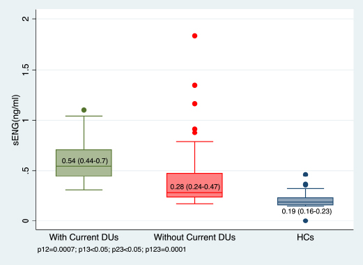
sENG levels in SSc patients with active DUs, SSc patients without active DUs, and healthy control sp12: Mann-Whitney test; p13,p23: T-test; p123: Kruskal-Wallis test.
Abbreviations: DU: digital ulcers, HC: healthy controls
A further subgroup analysis was performed among patients with active DUs (n = 30), those with a history of DUs but no active ulcers at enrollment (n = 10), and those who had never experienced DUs during the disease course (n = 20). The Kruskal-Wallis test showed a significant difference in sENG concentrations across these three groups. Patients with active DUs had significantly higher sENG concentrations than those with previous DUs but no active ulcers (median: 0.54 ng/mL; IQR: 0.44 to 0.70 versus 0.27 ng/mL; IQR: 0.21 to 0.45; p < 0.05) and those who had never had DUs (median: 0.32 ng/mL; IQR: 0.25 to 0.69; p = 0.009). In contrast, no significant difference was observed between patients with prior DUs and those without (p = 0.344). These findings are illustrated in Fig. (2).
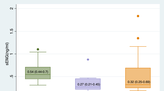
sENG levels among SSc patients who had never had DUs, those with previous DUs but no active ulcers, and those with active Dus p12, p13, p23: Mann-Whitney test; p123: Kruskal-Wallis test.
Abbreviations: DU: digital ulcers
Receiver operating characteristic curve analysis was conducted to evaluate the discriminative performance of sENG. An sENG cutoff value of 0.24 ng/mL differentiated SSc patients from healthy controls, with an area under the curve of 0.91 (p < 0.05), as shown in Fig. (3a). In addition, an sENG cutoff value of 0.43 ng/mL discriminated SSc patients with active DUs from those without active DUs, with an area under the curve of 0.75 (p < 0.0001), as shown in Fig. (3b).
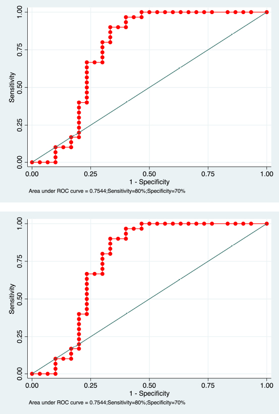
ROC curve analysis of sENG levels.
a. sENG level ≥ 0.24 ng/mL differentiated SSc patients from healthy controls.
b. sENG level ≥ 0.43 ng/mL was used for predicting DUs in SSc patients.
3.3. Clinical and Laboratory Correlation
Associations between clinical and laboratory characteristics and serum soluble endoglin (sENG) concentrations in patients with systemic sclerosis (SSc) are summarized in Table 2. Higher sENG concentrations were significantly associated with active or late nailfold capillaroscopy patterns (p < 0.001), elevated systolic pulmonary arterial pressure (p = 0.023), and a greater number of active digital ulcers (DUs) (p = 0.002). In contrast, no significant associations were observed between sENG concentrations and SSc subtype, interstitial lung disease on high-resolution computed tomography, impaired forced vital capacity, or the presence of anti-topoisomerase I, anti-centromere, and anti-RNA polymerase III antibodies (all p > 0.05).
Among patients with current DUs, the mean number of active DUs was 2.97 ± 2.02, with a range of 1 to 10 lesions. Spearman’s correlation analysis showed a significant positive correlation between sENG concentrations and the number of active DUs (rho = 0.39, p = 0.002), as illustrated in Fig. (4). However, sENG concentrations were not significantly correlated with DU severity, as assessed by the Digital Ulcer Clinical Assessment Score (rho = -0.09, p = 0.65; Table 2).
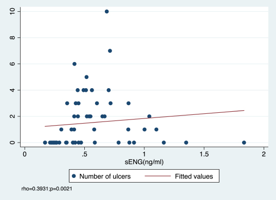
Correlation between sENG levels and the number of active DUs in SSc patients.
| Characteristics | sENG levels (ng/mL) | p | |
|---|---|---|---|
| Subtype | dcSSc (n=38). mean ± SD | 0.56 ± 0.35 | 0.55c |
| lcSSc (n=22). mean ± SD | 0.49 ± 0.26 | ||
| NC pattern | Early (n=17). mean ± SD | 0.31 ± 0.14 | <0.001c.* |
| Active/Late (n=43). mean ± SD | 0.62 ± 0.33 | ||
| ILD on HRCT | Yes (n=54). mean ± SD | 0.53 ± 0.31 | 0.87c |
| No (n=6). mean ± SD | 0.56 ± 0.41 | ||
| PAH | Yes (n=22) | 0.67 ± 0.38 | 0.023c.* |
| No (n=38) | 0.46 ± 0.26 | ||
| Impaired FVC | Yes (n=36) | 0.59 ± 0.36 | 0.178c |
| No (n=24) | 0.45 ± 0.23 | ||
| Anti-topoisomerase I ab | Yes (n=46). mean ± SD | 0.53 ± 0.28 | 0.52c |
| No (n=14). mean ± SD | 0.54 ± 0.43 | ||
| Anti-centromere ab | Yes (n=5). mean ± SD | 0.45 ± 0.13 | 0.88c |
| No (n=55). mean ± SD | 0.54 ± 0.33 | ||
| Anti-ARN polymerase III ab | Yes (n=2). mean ± SD | 0.7 ± 0.25 | 0.23c |
| No (n=58). mean ± SD | 0.53 ± 0.32 | ||
| Number of DUs | Rho = 0.39 | 0.002e.* | |
| DUCAS | Rho = -0.089 | 0.65e | |
Abbreviations: SD: standard, dcSSc: diffuse cutaneous systemic sclerosis, lcSSc: limited cutaneous systemic sclerosis, NC: nailfold capillaroscopy, ILD: interstitial lung disease, HRCT: high-resolution computed tomography, PAH: pulmonary arterial hypertension, FVC: forced vital capacity, ab: antibody, DU: digital ulcer, DUCAS: Digital Ulcer Clinical Assessment Score.
cMann-Whitney test
e Spearman’s rank correlation coefficient
*Statistical significance for a level of 5%
4. DISCUSSION
To our knowledge, this is the first study to evaluate serum soluble endoglin in relation to active digital ulcers in a Vietnamese systemic sclerosis cohort, and one of the few to distinguish active ulceration from previously healed ulcers and the absence of DU history. This distinction is clinically important because active DUs represent ongoing ischemic microvascular injury, whereas previous DUs may reflect remote vascular damage or long-term ulcer susceptibility. By separating these subgroups, our study provides a more refined interpretation of sENG as a biomarker of current vascular activity rather than DU history alone. The finding that sENG was elevated predominantly in patients with active DUs, rather than in those with previously healed ulcers, compared with DU-naïve patients, supports the hypothesis that circulating sENG may be linked to active endothelial injury and impaired vascular repair in SSc. These results add population-specific evidence from Southeast Asia and help address an important gap in the literature on vascular biomarkers for SSc-related digital ulcer disease.
This cross-sectional case-control study evaluated serum soluble endoglin as a potential vascular biomarker in Vietnamese patients with systemic sclerosis and demonstrated several key findings. Serum sENG levels were higher in patients with SSc than in healthy controls and increased stepwise across clinical groups, with median levels of 0.19 ng/mL in healthy controls, 0.28 ng/mL in SSc patients without active digital ulcers, and 0.54 ng/mL in those with active digital ulcers. This pattern suggests that sENG may reflect the extent of endothelial disturbance and peripheral vasculopathy in SSc. ROC analysis further showed good discrimination between SSc patients and healthy controls (AUC = 0.91 at a cut-off of 0.24 ng/mL) and moderate discrimination between SSc patients with and without active digital ulcers (AUC = 0.75 at a cut-off of 0.43 ng/mL). Notably, sENG was positively correlated with the number of active digital ulcers but not with DUCAS-assessed severity, suggesting that it may better capture active ulcer burden than composite ulcer complexity. Higher sENG levels were also associated with active or late nailfold capillaroscopy patterns and elevated systolic pulmo-nary arterial pressure, consistent with evidence linking sENG to the broader spectrum of SSc vasculopathy [1, 2].
The higher serum sENG levels observed in SSc patients compared with healthy controls in this study are biologically plausible given the role of endoglin in endothelial homeostasis and vascular remodeling. Endoglin, or CD105, is a transmembrane glycoprotein predominantly expressed on endothelial cells and acts as an accessory coreceptor in the TGFβ signaling pathway, thereby modulating angiogenesis, endothelial activation, and vascular repair (Grignaschi et al., 2022) [2]. Its soluble form is generated through proteolytic shedding of the membrane-bound ectodomain and may exert antiangiogenic effects by interfering with TGFβ-dependent endothelial nitric oxide synthase activation and impairing endothelial repair (Grignaschi et al., 2022) [2]. Because SSc is characterized by persistent endothelial injury, defective angiogenesis, and progressive capillary loss, elevated circulating sENG may reflect an imbalance in angiogenesis and ongoing endothelial dysfunction in SSc vasculopathy (Fioretto et al., 2023) [1]. Our finding is consistent with Wipff et al. (2008), who reported significantly higher sENG concentrations in 187 SSc patients than in age- and sex matched controls, with associations between sENG and vascular features, including cutaneous ulcerations and anticentromere antibody positivity [1, 11]. However, previous findings have not been entirely consistent, as summarized by Grignaschi et al. (2022) [2]. Differences in disease phenotype, sample size, ethnicity, treatment exposure, sample handling, and assay methodology may partly explain heterogeneity in circulating sENG measurements across cohorts [2].
A key finding of this study is that serum sENG levels were significantly higher in SSc patients with active digital ulcers than in those without active ulcers. More importantly, among patients without active DUs, sENG levels did not differ significantly between those with previous healed ulcers and those who had never experienced DUs. This distinction is clinically relevant because most previous studies classified patients broadly according to the presence or absence of DUs, without clearly separating active ulceration from healed or remote DU history [17]. Wipff et al. (2008) reported elevated sENG levels in SSc patients with cutaneous ulcerations [11], while Silva et al. (2016) found higher endoglin levels in patients with active DUs at baseline than in those without DU involvement until enrolment [18]. However, these studies did not specifically compare patients with active DUs, previous healed DUs, and no DU history as separate groups. The lack of difference between previous healed DUs and never DUs in our cohort suggests that sENG may be more closely related to ongoing ischemic microvascular injury than to remote ulcer susceptibility alone. This interpretation is consistent with the concept that circulating markers of endothelial injury may vary with vascular disease activity rather than representing fixed historical damage (Grignaschi et al., 2022) [2]. Nevertheless, whether sENG levels decline after DU healing or predict recurrence remains to be confirmed in longitudinal studies.
In this study, serum sENG was positively correlated with the number of active digital ulcers (Spearman’s rho = 0.39, p = 0.002), suggesting that higher circulating sENG levels may reflect greater active ulcer burden and more extensive ongoing ischemic microvascular injury. This association is biologically plausible because multiple concurrent ulcers may indicate broader endothelial disruption and more pronounced angiogenic imbalance, potentially increasing the release of soluble endoglin. In contrast, sENG was not significantly correlated with DU severity assessed by the Digital Ulcer Clinical Assessment Score (DUCAS; rho = -0.09, p = 0.65). This difference may be explained by the multidimensional nature of DUCAS, which incorporates not only ulcer number but also new ulceration, gangrene, infection, surgical intervention, unscheduled hospitalization, and analgesic use (Bruni et al., 2019) [9]. These components may be influenced by wound care, timing of assessment, pain management, infection control, healthcare access, and hospitalization practices, rather than endothelial injury alone. Therefore, DUCAS may better capture the overall clinical and functional burden of active DUs, whereas sENG may more specifically reflect the biological activity of microvascular injury (Fioretto et al., 2023) [1]. Taken together, these findings suggest that sENG and DUCAS provide complementary information and may be considered together when characterizing active DU disease in SSc.
ROC analysis showed that serum sENG had good to excellent discriminative performance for distinguishing SSc patients from healthy controls, with an AUC of 0.91 at an optimal cut-off of 0.24 ng/mL. For differentiate SSc patients with active digital ulcers from those without, sENG showed moderate discrimination, with an AUC of 0.75 at a cut-off of 0.43 ng/mL. These findings suggest that sENG may have adjunctive value in identifying SSc-related vascular involvement and active DU-related vasculopathy, although it should not be considered a standalone diagnostic test [19]. This interpretation is consistent with previous literature supporting elevated circulating sENG as a marker of SSc vascular involvement and its association with the digital ulcer phenotype, as reviewed by Grignaschi et al. (2023) and Bruni et al. (2022) [2, 9]. The cut-off values in our study were lower than those reported by Wipff et al. (2008), who described markedly elevated sENG levels in a European SSc cohort [11], and lower than the threshold of greater than 4.215 ng/mL associated with digital ulcer tendency reported by Silva et al. (2015) [20]. However, they were closer to the cutoff of 0.29 ng/mL, with an AUC of 0.748, for active disease flare, as identified in a recent study using a comparable low-range ELISA platform. These discrepancies should be interpreted cautiously, as they may reflect differences in ELISA assay characteristics, antibody calibration, sample handling, disease duration, treatment exposure, DU definition, disease subtype distribution, and ethnic variation across European, Latin American, and Southeast Asian populations. Coral Alvarado et al. (2010) also reported detectable sENG elevation at comparatively modest absolute concentrations in a Latin American cohort, further emphasizing the context-dependent nature of absolute thresholds [21]. Overall, sENG may support clinical assessment of SSc-associated vasculopathy as an adjunctive biomarker, but these cutoff values require prospective, population-specific, and assay-specific external validation before routine clinical application.
CONCLUSION
Soluble endoglin levels were elevated in SSc patients with DUs, supporting the potential role of endoglin in SSc-related vasculopathy. Increased sENG concentrations were particularly associated with active DUs and a higher number of ulcerative lesions, suggesting that sENG may have predictive value for the development and progression of digital ulcers. Further multicenter studies with larger populations and longitudinal follow-up are needed to validate sENG as a biomarker for SSc-related DUs. In addition, future research should clarify the distinct roles of different ENG isoforms, particularly short and long ENG, in the pathogenesis of SSc vasculopathy. These findings may contribute to the development of new diagnostic, prognostic, and potentially therapeutic approaches for patients with SSc.
STUDY LIMITATIONS
This study has several limitations. First, the relatively small sample size and single-center recruitment may limit the generalizability of the findings to broader SSc populations. Second, the cross-sectional case-control design precluded assessment of temporal relationships and did not allow us to determine whether elevated serum sENG levels predict the subsequent development, healing, or recurrence of digital ulcers. Third, only circulating sENG concentrations were measured, without parallel assessment of ENG expression in vascular tissues, which limited mechanistic interpretation. Finally, digital ulcer development is likely driven by complex interactions among vascular, inflammatory, immune, and angiogenic pathways; therefore, sENG should not be interpreted as a standalone marker. Larger multicenter longitudinal studies incorporating repeated sENG measurements and broader biomarker panels are needed to validate these findings and clarify the clinical relevance of sENG in SSc-related digital ulcer disease.
AUTHORS’ CONTRIBUTIONS
The authors confirm their contributions to the paper as follows: H.T. N., H.T.T. Do, and D.H. Le.: Contributed substantially to the conception and design of the study; H.T. N., V.T.H. N., P.T. H., and G.T.H. Q.: Contributed to data collection; H.T. N. and M.H. Le.: Contributed to data acquisition, analysis, and interpretation; H.T. N., H.T.T. Do., and D.H. Le.: Drafted the manuscript and revised it critically for important intellectual content. All authors approved the final version of the manuscript for publication and agree to be accountable for all aspects of the work, including the accuracy and integrity of any part of the study.
LIST OF ABBREVIATIONS
| AUC | = Area under the curve |
| DUs | = Digital ulcers |
| DUCAS | = Digital ulcer clinical assessment score |
| ELISA | = Enzyme-linked immunosorbent assay |
| HRCT | = High-resolution computed tomography |
| ILD | = Interstitial lung disease |
| IQR | = Interquartile range |
| NVC | = Nailfold capillaroscopy |
| ROC | = Receiver operating characteristic |
| sENG | = Soluble endoglin |
| SSc | = Systemic sclerosis |
| sPAP | = Systolic pulmonary arterial pressure |
ETHICS APPROVAL AND CONSENT TO PARTICIPATE
This study was approved by the Hanoi Medical University Institutional Review Board, Vietnam under approval number 1343/GCN-HMUIRB on April 04, 2024.
HUMAN AND ANIMAL RIGHTS
All procedures involving human participants were conducted in accordance with the ethical standards of the committee responsible for human experimentation (institutional and national), and with the Helsinki Declaration of 1975, as revised in 2013.
CONSENT FOR PUBLICATION
Written informed consent was obtained from all individual participants prior to enrollment in the study, including consent for data collection and blood sample collection.
AVAILABILITY OF DATA AND MATERIALS
The data of current study are available from corresponding author, [D.H.L], on a reasonable request.
ACKNOWLEDGEMENTS
The authors sincerely thank the Autoimmune Connective Tissue Disease Department for facilitating patient recruitment and the Department of Laboratory Medicine for their support with ELISA testing. The authors also thank Tung S. Tran, MD, for his statistical support.