All published articles of this journal are available on ScienceDirect.

Recent Insights On Diabetic Dermopathy
Abstract
Background:
Diabetic dermopathy consists of small, round, brown atrophic skin lesions that occur on the shins of patients with diabetes. Its proper diagnosis is essential for proper management.
Objective:
The present study has been undertaken to study the complications, signs, symptoms, prevention and cure of dermopathy caused by diabetes.
Material and Methods:
Dermopathy was studied in brief with the help of literature available in the form of articles, various databases, medical news, etc.
Result:
Proper diagnosis and cure are necessary at early stages to prevent future complications associated with it.
Conclusion:
Diabetic dermopathy requires no treatment, but may be a surrogate for more serious complications of diabetes, which require investigation and management.
1. INTRODUCTION
Diabetic dermopathy is the commonest skin condition that happens in patients with Diabetes Mellitus. It is also known as pigmented pretibial patches, spotted leg syndrome or diabetic dermangiopathy or shin spots. It is found in up to five-hundredths of diabetics and is the most typical connective tissue finding in patients with Diabetes Mellitus. The condition was initially rumored in 1964 by Melin, circumscribed, brown symptoms, skin lesions occurring on the lower extremities [1]. Within the literature, there is restricted knowledge relating to early-stage skin disorders in Diabetic Mellitus patients, particularly specializing in non-injured skin. Higher understanding of the burden of skin disorders in polygenic disorder patients could raise awareness on hindrance and its management [2]. Regarding connective tissue infections, fungus etiology seems to be foremost common in people with micro-organism origin area [3-5]. Diabetic dermopathy presents brown or pink well-demarcated macules or topped papules on the bilateral pretibial areas of patients with Diabetes Mellitus. Typically, the lesions area are unit spherical or oval and are 1cm in diameter, however larger patches area unit are often seen. The surface is usually slightly depressed with a pointy drop off from traditional adjacent skin. The arrangement of lesions is usually sorted and sometimes linear. No pain or itching is associated [6, 7]. The signs and complications of Diabetic dermopathy need to be correctly monitored for its proper hindrance and cure. This critique focuses on the varied aspects of Diabetic dermopathy. Diabetic dermopathy is the most prevailing skin presentation together with hyperpigmented and symptom macules or papules with no sharp border and frequently occurring on the front leg in polygenic disorder patients. The designation of Diabetic dermopathy is clinical and is formed by description and physical examination. The prevalence of scars and hyperpigmented, symptom spots with sharp borders is observed Diabetic dermopathy has reported 9%-55% rife and is probably going to be additional rife within the patients over fifty years and with longer length of polygenic disorders. Meanwhile, it happens sometimes before diabetic retinopathy and kidney diseases [8]. The present review article deals with the complications, signs & symptoms, prevention and treatment of Diabetic dermopathy.
2. COMPLICATIONS
The incidence of Diabetic dermopathy will increase with age. It is generally seen in patients aged older than fifty. Men show associate inflated incidences as compared to ladies. Though situated bilaterally, their distribution is uneven. Lesions don't itch or cause pain. Management of glucose levels doesn't have an effect on the result of the lesions. There is no correlation between diabetic dermopathy and fat or high blood pressure.The designation of diabetic dermopathy is very important owing to its association with diabetic microangiopathic complications together with retinopathy, neuropathy, and kidney diseases. The presence of diabetic dermopathy could also be an associated indicator of alternative additional serious pathology and its prevalence has been related to each microvascular complications and enormous vessel malady [9]. In Melin’s original study of sixty nine patients with skin lesions had retinopathy; solely twenty fifth of the cluster while not skin lesions had retinopathy [10]. These patches could also be oval or circular. Some individuals mistake them for age spots. This disorder most frequently happens on the front of eachleg. However, the legs might not be affected to a similar degree [11]. The patches don't hurt, open upor itch. Hyperpigmented macules on the shins, alleged diabetic dermopathy has been termed the foremost common cutaneous finding in polygenic disorder. It is sometimes noted as an irregular basis spherical or oval, circumscribed, shallow lesions varied in range from few to several, that are sometimes bilateral however not symmetrically distributed [12, 13]. The presence of many hyperpigmented symptom macules on the shins is alleged to be a comparatively common finding in patients with polygenic disorder. Skin lesions in patients with polygenic disorder, notably on their feet are common and complex in nature [14, 15]. Diabetic dermopathy can usually appear as circular, scaly, brown patches. It is most typically seen on the anterior side of the bilateral lower extremities [16, 17].Skin atrophy occurs from microangiopathy.Decreased blood ensures macroangiopathy. Sensory and involuntary pathology, leading to abnormal blood distribution that will cause bone demineralization and Charcot's joint [18]. Although skin blood flow at basal vital sign isn't completely different from that in nondiabetic subjects, there's a four-hundredth to five hundredth reduction of heat-stimulated flow in diabetic patients compared with the nondiabetic management population [19]. Heat stimulation elicits supreme dilation of the body covering microvasculature, so the reduced flow values in polygenic disorder indicate a loss of skin blood flow reserve. This impairment of blood flow reserve affects patients with one and two polygenic disorder and presumptively reflects a diabetic body covering microangiopathy [20, 21].
3. SIGNS & SYMPTOMS OF DIABETIC DERMOPATHY
Diabetic dermopathy seems as tiny, circumscribed, brown symptom skin lesions but one cm in diameter though some is also elongated and up to 5 cm. Lesions begin as non-blanching, scaly, red or purple, spherical or oval macules or papules. There is also sclerosis with a central depression or organic process. These lesions afterward change to the characteristic scar-like lesions of diabetic dermopathy. The intensity of pigmentation corresponds to the degree of atrophy with the darkest lesions additionally being the foremost symptom. Lesions last for on average eighteen to twenty-four months before attenuation to minimal symptom macules or clearing utterly. In some cases, the brown color disappears and is replaced by a small coloration. As older lesions are clear, new lesions seem to appear. The patches are slightly scaly and are typically spherical or oval. The lesions don't unremarkably burn or sting even if they seem as if they could feel painful. Diabetic dermopathy in all probability represents post-traumatic atrophy and post inflammatory physiological state in poorly vascularized skin. Recent reports showed that the majority of patients have a rise in glycosylated hemoprotein and an extended history of the polygenic disorder. It is double as common in men as in girls. These patches are found in traditional old individuals. Diabetic dermopathy happens on the shins in an exceedingly bilateral asymmetrical distribution. It is common in patients over the age of fifty. The patches are slightly scaly and are typically spherical or oval. Long-standing patches might become faintly indented.
Locations of diabetic dermopathy are:
Skin alterations because of diabetic complications:
- Diabetic foot
- Cutaneous infections related to polygenic disease
- furunculosis
- Erythrasma
- Xanthomatosis
- Xanthelasma
- Pycomycetes
- Malignant inflammation media [24-27]
- Spots or lesions on the shins of an individual with Diabetes Mellitus.
- The lesions as shown in Fig. (1), or spots can even be found on the front a part of the thighs, scalp, sides of the feet, trunk, and forearm.
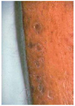 Fig. (1). Macro photographs of diabetic dermopathy lesions
 - The spots are bilateral, found on each shins at constant time.
- Over time, the patch sounds like associate degree age spot [28-30].
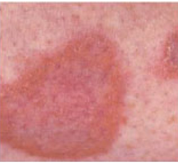
4. SKIN CHANGES IN DIABETIC GANGRENE
It is a condition that occurs when body tissue dies as shown in Fig (2). It is caused by a loss of blood supply due to an underlying illness, injury, and/or infection. Fingers, toes, and limbs are most often affected, but gangrene can also occur inside the body, damaging organs and muscles. Blood transports oxygen and nutrients throughout the body to feed cells, it delivers disease-fighting antibodies that protects the body from infection. When blood cannot travel freely throughout the body, cells cannot survive, infection can develop and tissue can die from gangrene [31, 32].
5. SKIN CHANGES IN DIABETIC SCLERODERMA
While rare, this skin problem affects people with type 2 diabetes, causing a thickening of the skin on the back of the neck and upper back. The treatment is to bring blood sugar level under monitoring. Lotions and moisturizers may help to soften skin. Scleroderma-like skin changes as a part of “diabetic hand limited joint mobility syndrome” is a clinical entity reported in 10-50% of adolescents and adults with diabetes mellitus. The skin changes may occur early in the disease. The assumption is that vessel and connective tissue alterations, as well as the impairment of the immune system and other associated metabolic changes caused by diabetes, play an important role [33, 34].
6. SKIN CHANGES IN NECROBIOSIS LIPOIDICA
Necrobiosis lipoidica is a necrotizing skin condition that usually occurs in patients with diabetes mellitus. In the former case it may be called Necrobiosis Lipoidica Diabeticorum (NLD).NLD occurs in approximately 0.3% of the diabetic population, with the majority of sufferers being women approximately 3:1 females to males affected. The severity or control of diabetes in an individual does not affect who will or will not get NLD [35-37].
7. PREVENTION
Since diabetic dermopathy is rife and customarily takes place before retinopathy and uropathy, early diagnosing of dermopathy might facilitate to predict the incidences of retinopathy and uropathy, delay their incidence and adverse and dangerous consequences or prevent their progression by managing polygenic disease and alternative factors additional expeditiously if the association between dermopathy and incidence of retinopathy and uropathy is confirmed within the patients with sort two polygenic disease. Monitoring of blood glucose level regularly is essential. Prevention of any sorts of injury or infection can be done by keeping skin moisturized. Changes on skin should be discussed with doctor. Proper healthy, sugar free diet at regular intervals is essential [38-40]. If the doctor prescribes medication, staying with it facilitates the integrity of skin. There are lots of ways to manage it. To prevent complications, blood glucose level should be managed by maintaining a healthy diet, exercise often and the medications prescribed by doctor [41-43].
8. TREATMENT
Diabetic dermopathy typically improves as time passes by. Skin should be kept healthy and moisturized. We should avoid any varieties of injury to the legs therefore on forestall the lesions or spots from more progression. There is very no need for medical intervention once it involves lesions, spots, or patches secondary to polygenic disease. However, it's necessary to regulate blood glucose level as a result of it's the amount one triggering issue for dermopathy [44-46]. Treatment for diabetic dermopathy is all regarding taking excellent care of blood glucose level [46-48]. Once blood glucose level is controlled, it's not solely the diabetic dermopathy that may be prevented however still different complications of polygenic disease. Home remedies for diabetic dermopathy are all regarding ingestion of a healthy diet, healthy and safe exercise routines. Another necessary issue is to stay skin healthy, particularly within the areas wherever spots or patches presumably to occur [49-58]. No treatment is usually recommended or has been shown to be effective forthe lesions of diabetic dermopathy. Cosmetic camouflage is used to disguise the looks of the skin lesions if needed [59-68].
CONCLUSION
From the above observations it is clear that skin is concerned in diabetics very often. Diabetic dermopathy lesions or shin spots are harmless. They typically don't need any treatment and have a tendency to travel away once a number of years, significantly following improved glucose management. Whereas if any corpulent patient present with multiple shin spots having fast glucose levels towards the upper facet of traditional in conjunction with the a positive case history of diabetes, more investigation should be conducted to rule out the likelihood of early sickness. Ingestion of a healthy diet, healthy and safe exercise routines and medicines prescribed by the physician are all meant to be done in diabetic dermopathy.
CONSENT FOR PUBLICATION
Not applicable.
CONFLICT OF INTEREST
The authors declare no conflict of interest, financial or otherwise.
ACKNOWLEDGEMENTS
Declared none.

